
Understanding Cracked Tooth Syndrome
Cracked tooth syndrome (CTS) describes a spectrum of incomplete tooth fractures that cause pain on biting and/or cold sensitivity but do not yet show radiographic evidence of complete fracture or pulp necrosis. The crack typically runs vertically or obliquely through the coronal tooth structure — often initiating at the base of a cusp under large amalgam restorations or from occlusal stress in heavily restored teeth.
CTS pain has a distinctive character: sharp, momentary, provoked specifically by biting on a particular spot or releasing bite pressure. This "rebound" pain is caused by the hydrodynamic fluid movement in the dentin tubules as the crack flexes open and snaps closed under load. As the crack deepens, pulpal inflammation (pulpitis) develops, and the pain transitions from sharp-on-biting to lingering, spontaneous, and eventually constant — the progression toward irreversible pulpitis and the need for root canal treatment.
Per tooth, fee-for-service cash pay: the US fee range for a porcelain onlay (Pearl, from ADA fee survey data, updated May 2026). It does not price crack arrest or the full adhesive protocol separately, so ask the clinic for a written estimate. For comparison, root canal treatment on a molar costs $833–$2,171 (CareCredit, September 2026) and a porcelain crown after it $1,015–$2,670 (Pearl, updated May 2026) — the treatment the crack cascade leads to if left untreated.
Why Conventional Crowns Fail to Arrest Cracks
The conventional response to CTS is full-coverage crown placement. The rationale is sound in theory: a circumferential crown "hoops" the tooth and prevents the cracked cusps from flexing apart under load. In practice, however, crown preparation has a critical failure mode: the preparation bur must cut across the crack line during axial reduction, opening the crack to the oral environment and potentially seeding bacteria into the fracture before the crown is cemented.
More fundamentally, a conventionally cemented crown relies on mechanical retention — it does not seal and bind the crack the way an adhesive restoration does. If the crack extends below the crown margin (into the root), the crown provides no structural support below its apical extent. Adhesive restorations, by contrast, bond to the crack surfaces and resist their separation under load — not just by covering them, but by chemically joining them.
The Biomimetic Crack Arrest Protocol
Diagnosis and Crack Mapping
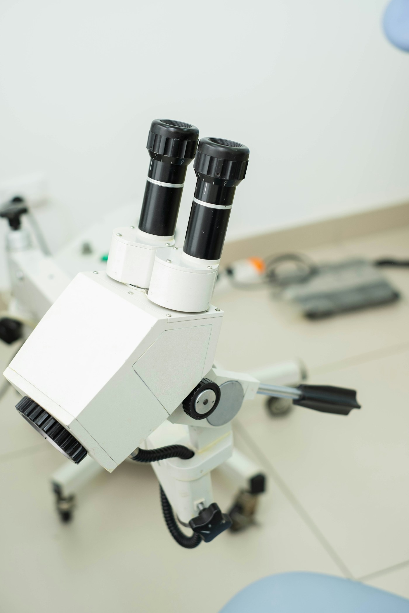
Accurate crack diagnosis requires transillumination, dye staining (crystal violet or methylene blue), and bite test localization with a Tooth Slooth or Fractfinder. The crack must be mapped in three dimensions: direction, depth relative to the pulp, and extent on the root surface (if any). A crack that has extended into the root below the crestal bone is a contraindication to biomimetic restoration — the tooth requires extraction or intentional replantation.
Preparation and Crack Removal
The preparation removes the undermined and cracked tooth structure while preserving all sound cusp and axial wall anatomy. The preparation is designed to expose the full crack extent so it can be sealed — the crack should not be left beneath the preparation floor. Under transillumination or methylene blue restaining after preparation, the crack extent is verified to be within the preparation boundary.
Immediate Dentin Sealing and Crack Binding
Immediately after preparation, the exposed dentin and crack surfaces are treated with Immediate Dentin Sealing. The adhesive resin penetrates the crack by capillary action, impregnating the crack walls and creating a resin "pin" along the crack's length. When light-cured, this resin effectively seals the crack against bacterial and fluid penetration — the primary mechanism of ongoing pulpal irritation. This is the step that converts a progressive fracture into a stable, sealed, biologically inert structural feature.
Stress-Reduced Composite Base
A low-modulus, stress-absorbing composite base is placed over the deeper portions of the preparation, providing a cushioning layer between the ceramic restoration and the underlying dentin. This base distributes occlusal load more evenly across the crack surfaces, reducing the peak stress concentration at the crack tip that would otherwise drive further propagation.
Ceramic Onlay or Overlay Cementation
The indirect ceramic restoration selected depends on crack geometry. If one cusp is cracked, a cusp-replacing onlay is appropriate. If multiple cusps are involved or the crack runs through the central groove into both mesial and distal cusps, a full-occlusal ceramic overlay "hoops" the entire tooth simultaneously — replicating the mechanical principle of the circumferential crown without requiring axial wall reduction.
The ceramic is adhesively bonded at cementation using dual-cure resin cement, creating a restoration that is not merely retained on the tooth but is structurally fused to it — resisting crack separation at the adhesive interface in addition to the IDS layer within the crack itself.
The Critical Timing Variable
Biomimetic crack arrest is most predictable when performed before irreversible pulpitis develops. Once the patient reports lingering pain after a thermal stimulus lasting more than 5–10 seconds, spontaneous pain episodes, or pain that wakes them from sleep, the pulp is likely already irreversibly inflamed and root canal treatment is required regardless of the restorative approach. Early treatment is decisive.
Ribbond Fiber as a Crack Arrest Adjunct
In teeth with extensive crack networks or particularly high occlusal loads, biomimetic dentists may incorporate Ribbond ultrahigh-molecular-weight polyethylene fiber into the stress-reduced composite base. The fiber acts as a crack arrest mesh — replicating the collagen architecture of natural dentin in distributing tensile stress away from crack tips. See the full guide to Ribbond fiber reinforcement.
Post-Treatment Outcomes
Pain on biting typically resolves within days to two weeks of biomimetic onlay cementation as the sealed crack loses its hydrodynamic activity and the restored cusps are held from flexing apart under load. Residual cold sensitivity lasting 4–8 weeks is within normal range as the pulp recovers from ongoing irritation; this resolves without intervention in the majority of cases when pulpitis is still in the reversible phase at treatment time.
Long-term outcome data on biomimetic crack arrest restorations shows pulp survival rates of 80–90% at 5 years in teeth presenting with reversible pulpitis at baseline. This compares favorably to the 15–20% 10-year root canal rate for conventionally crowned teeth without specific crack management.
Credentials to Verify
- AOBMDAcademy of Biomimetic Dentistry Fellowship — cracked tooth management is a specific competency area within the AOBMD fellowship curriculum, requiring documented cases with crack diagnosis, IDS execution, and stress-reduced composite basing.
- ALLEMANAlleman Center Six Lessons — Lesson 3 and 4 specifically address cracked tooth management, pulp capping, and the adhesive protocol adaptations for teeth with compromised pulpal health.
Frequently Asked Questions
Can a cracked tooth heal on its own?
No. Unlike bone, tooth enamel and dentin have no regenerative capacity. A crack does not heal — it only propagates under continued functional load. The question is not whether to treat it, but when. Early treatment (reversible pulpitis, crack confined to coronal tooth structure) has the best prognosis.
Why did my dentist say the only option is a crown or root canal?
Crown placement is the conventional standard of care for CTS. Root canal treatment is recommended when pulpitis has progressed to irreversible. Biomimetic crack arrest as a specific protocol — using adhesive resin to seal and bind the crack before ceramic placement — is not universally taught in dental school and requires post-doctoral training to execute correctly. A second opinion from an AOBMD-credentialed dentist is warranted if you are in the early stages of CTS.
How do I know if my tooth is cracked?
Sharp, momentary pain provoked specifically by biting on one spot, or sharp pain on releasing bite pressure, is the most specific symptom. Cold sensitivity that lingers beyond 5 seconds suggests the pulp is involved. Your dentist can use a bite stick, transillumination, and dye staining to confirm and map the crack. CBCT imaging can sometimes visualize crack extent below the gumline.